A Leaf Under A Microscope
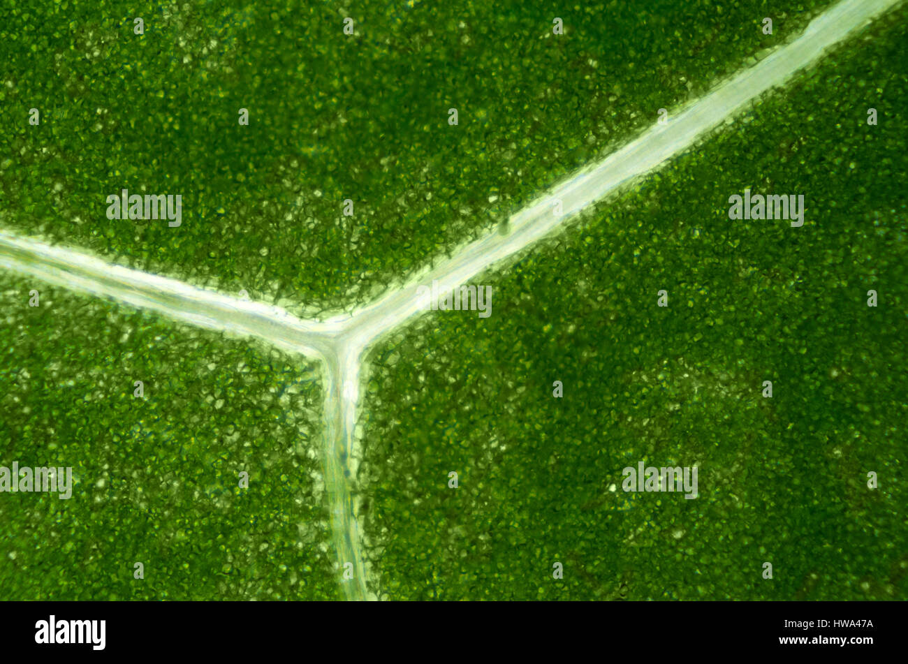
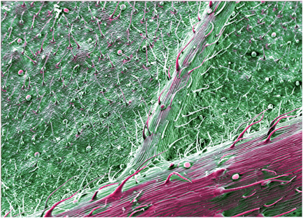
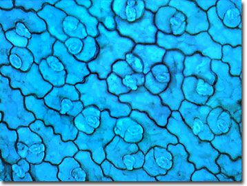
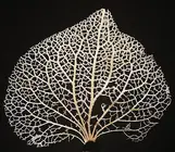
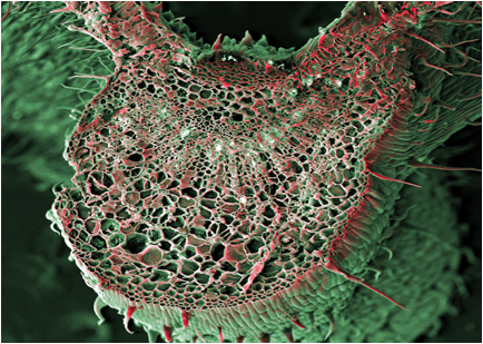
They're actually called Smiley Bundles or Vascular Bundles, which help to transport water, minerals and food.
A leaf under a microscope. Add 2 or 3 drops of salt solution to the leaf and replace the cover slip. Carefully peel off the glue. Look through the eyepiece and focus the leaf into view.
Observe the slide, first, under the lower magnification (i.e., 10 X) of a compound microscope and then, under the higher magnification (i.e., 45 X). If the student can see only a thick, dark mass when looking under the microscope, the slice is too thick. A Visual Exploration of Medicinal Sativa and C.
Identify the spongy and palisade parenchyma, and the size and location of the stomata. How to Record Microscope Observations In the field of science, recording observations while performing an experiment is one of the most useful tools available. Early scientists often kept very detailed journals of the experiments they performed, making entries for each individual experiment and writing down virtually.
What is moving in this leaf under a microscope?. Cover with a slip. When put under a microscope objects such as bananas, toothbrushes and even used dental floss look incredible.
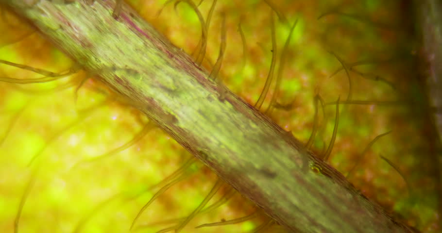
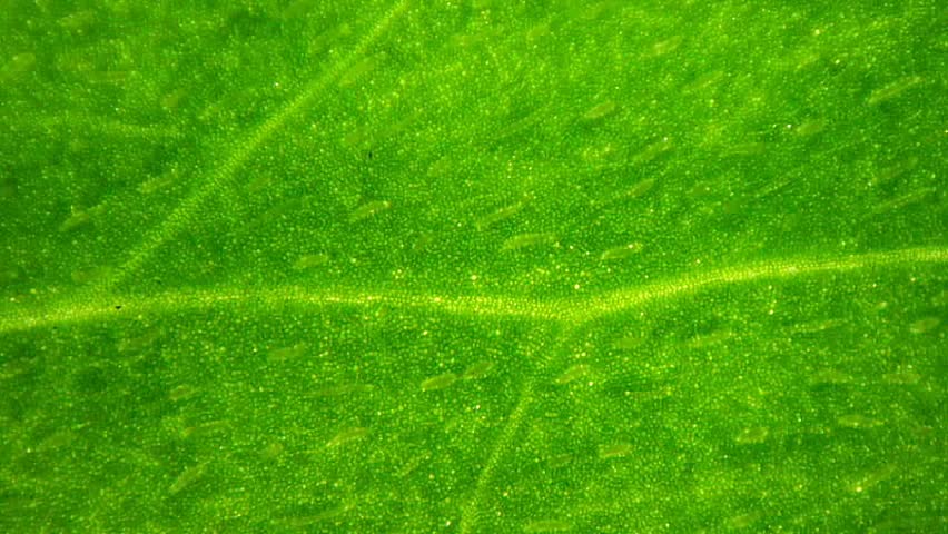
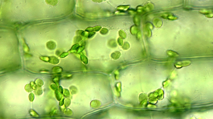
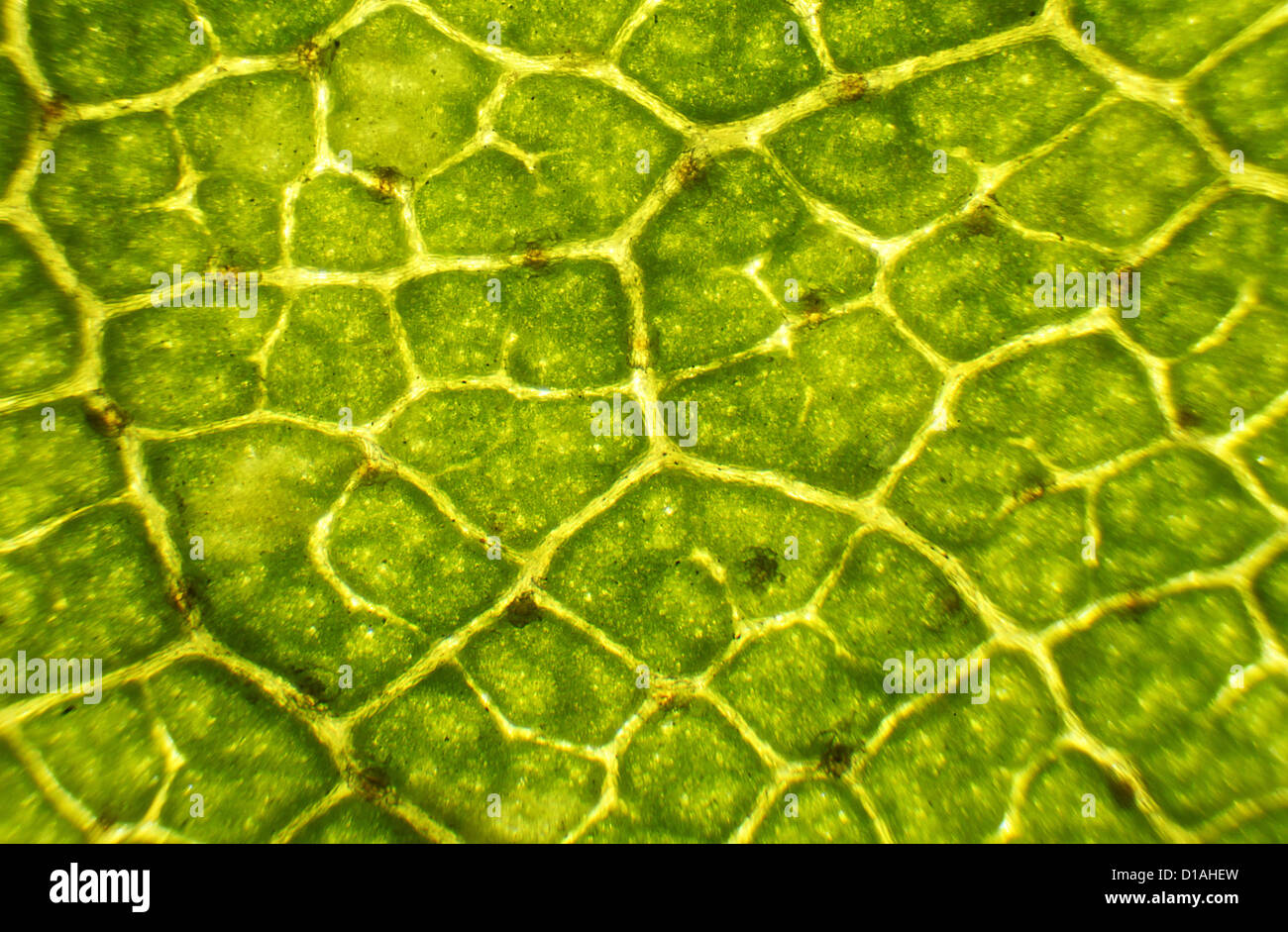
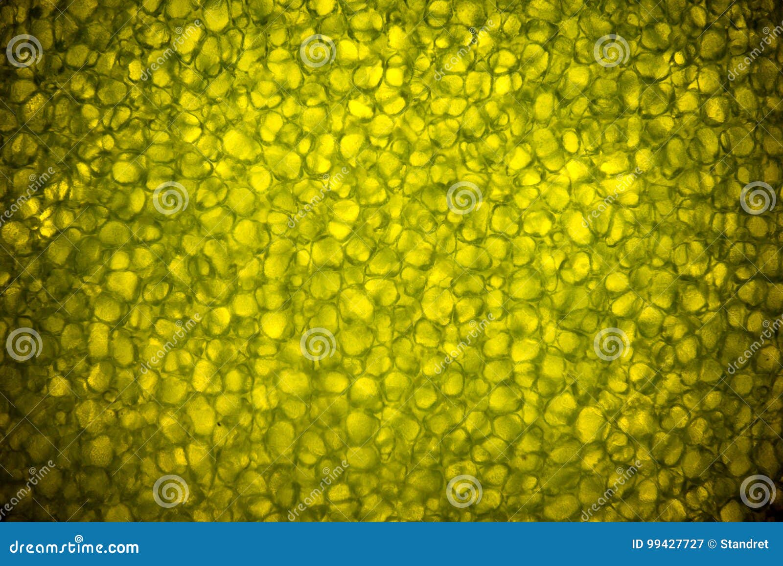
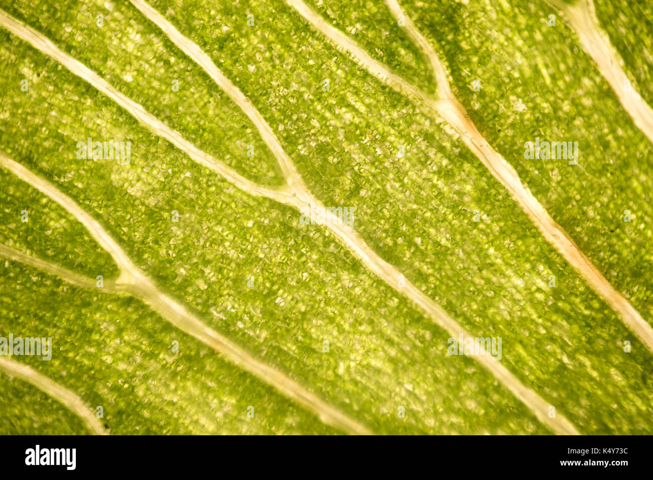
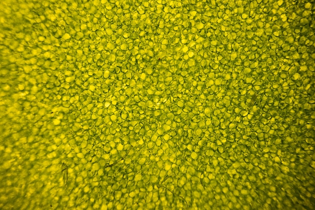
Have a peek below:. Micrograph, leaf under a microscope, organ-producing oxygen and carbon dioxide, the process of photosynthesis Coloured SEM of leaf mining fly (Agromyzidae) Macro texture of autumn maple leaf in vertical frame. Plant cell surface of leaf under microscope Sunflower leaf under the microscope Leaf cells under microscope.
Elodea Leaf Under Microscope Labeled. It has become transparent. Place under the microscope for observation.
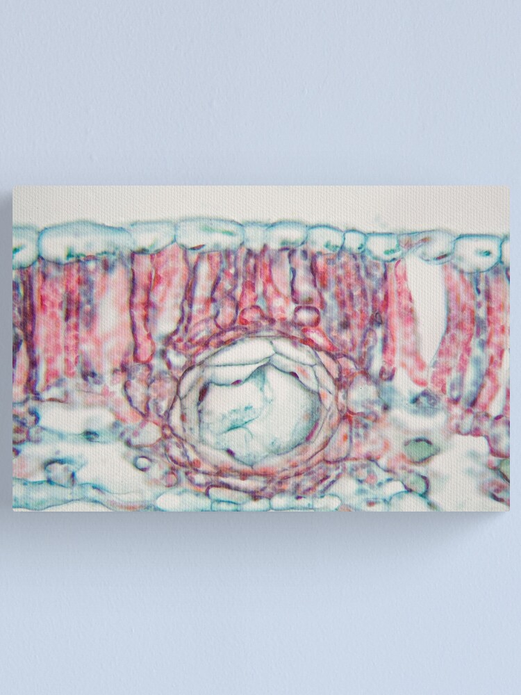
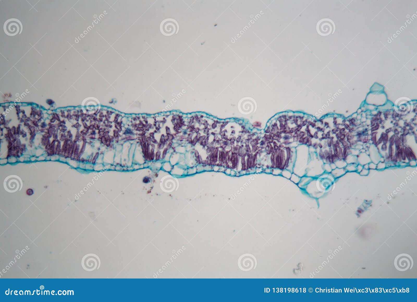
The light intensity must be sufficiently high to pass through the leaf. Dicot Leaf Cross Section (Dorsiventral Leaf) (Anatomical Structure of a Dicot Leaf- Ixora, Mangifera, Hibiscus) Ø Leaves are structurally well adapted to perform the photosynthesis, transpiration and gaseous exchange. Examine a prepared slide of an Oleander leaf.
Elodea leaf cell ilration from a microscope slide drop of 10 mackenzie zimbrick lab 3 labelling an elodea cell lab manual exercise 1 elodea leaf 400x general biology lab loyola university chicago. Sometimes the most mundane thing suddenly becomes mind-blowingly beautiful. Presence of very tiny black structures (pycnidia) in the center of these spots confirms they are symptoms of Septoria leaf spot.
(a) chenopodium leaf surface showing intact epidermal cells and salt glands;. Under a microscope, even the plainest items prove to be unbelievably complicated. Remove the cover slip and remove the water from the leaf with a paper towel.
This is actually not a microscope image, but was taken with the Canon PowerShot SX40 and Raynox macro conversion lens. Look at fresh Salvia under the dissecting microscope. Next, put the stage clips on to fasten it securely.
So yeah, you could say there's more to. Leaf Epidermis Stomata under microscope. 🔴 Subscribe so you don't miss any video!.
The total number of cells in each stage of the cell cycle is recorded and presented in the table below. In this case, the ovary is already well-developed and is good for examining under a scientific microscope. There is a ficus tree right outside of my classroom door, so that is what I use every year.
Wait several hours or overnight for the glue to dry. Look at the slide under the microscope to be sure the slide shows the cells clearly. How to Detect Moldy Buds To find out if your weed is moldy, first, get a lab analysis from your supplier.
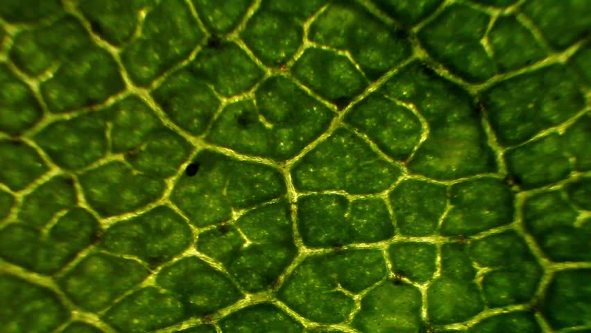
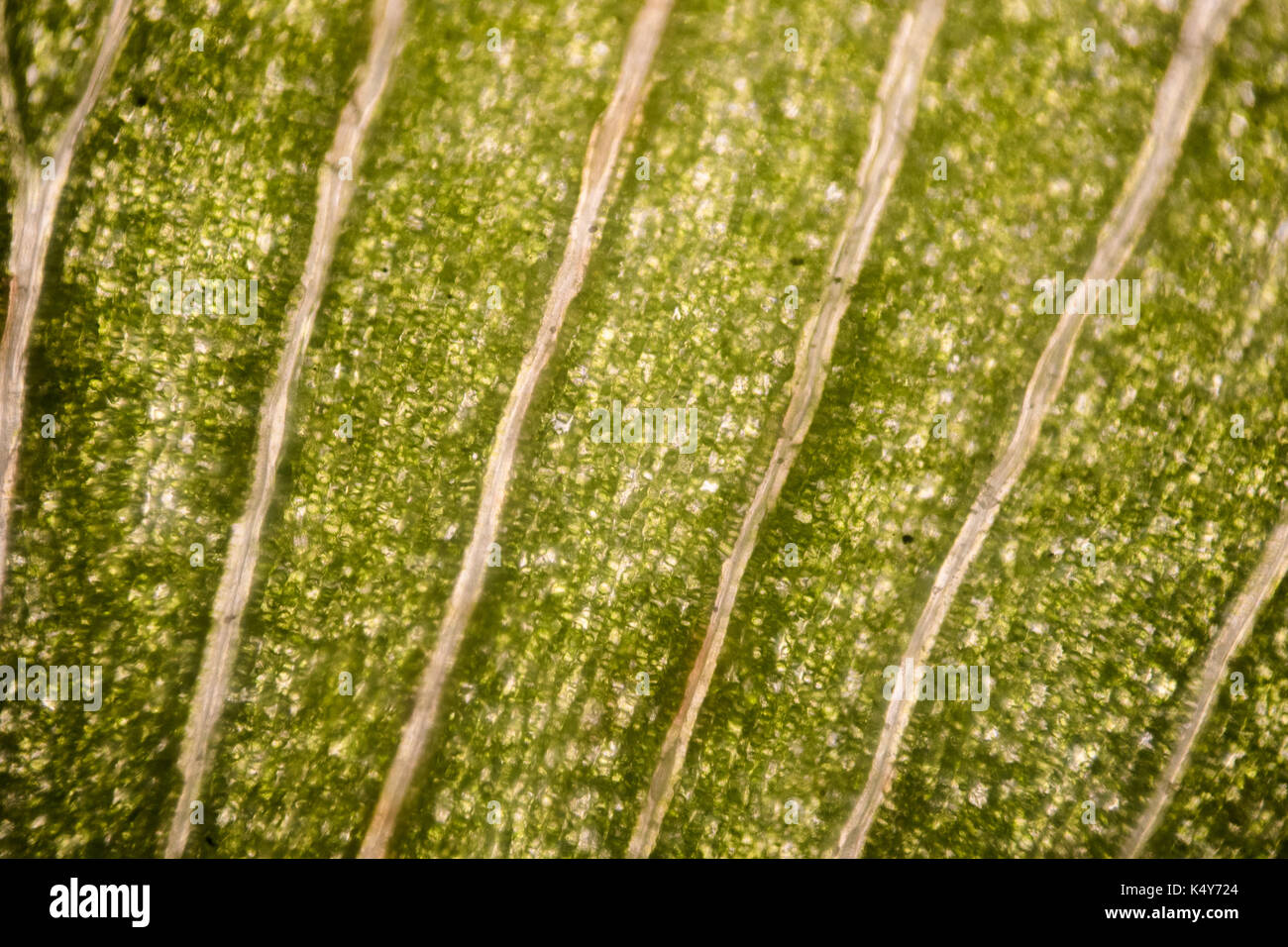
60x Cotton leaf under the microscope. The image represents a section of the leaf approximately 3 mm across. The cells look happy.
Elodea Leaf Microscope Lab;. Jan 27, 14 - Explore Michael Hanophy's board "Plant Anatomy I - Leaf, Stem, Root", followed by 239 people on Pinterest. The plant is native to the cool and warm waters of the Old World in.
When viewed with a microscope, they often look like coffee beans. Elodea Leaf Microscope Labeled;. Identify the xylem and phloem in the leaf.
Notice the upper and lower epidermis and location of the stomata. Scanning electron microscope image of cannabis budCannabis Under the Microscope:. These are the egg cells which will later develop into seeds.
Add the stain as you like and glycerine too. See more ideas about Root, Plants, Stem. Get premium, high resolution news photos at Getty Images.
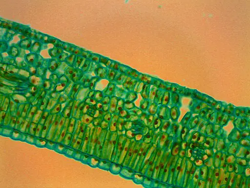
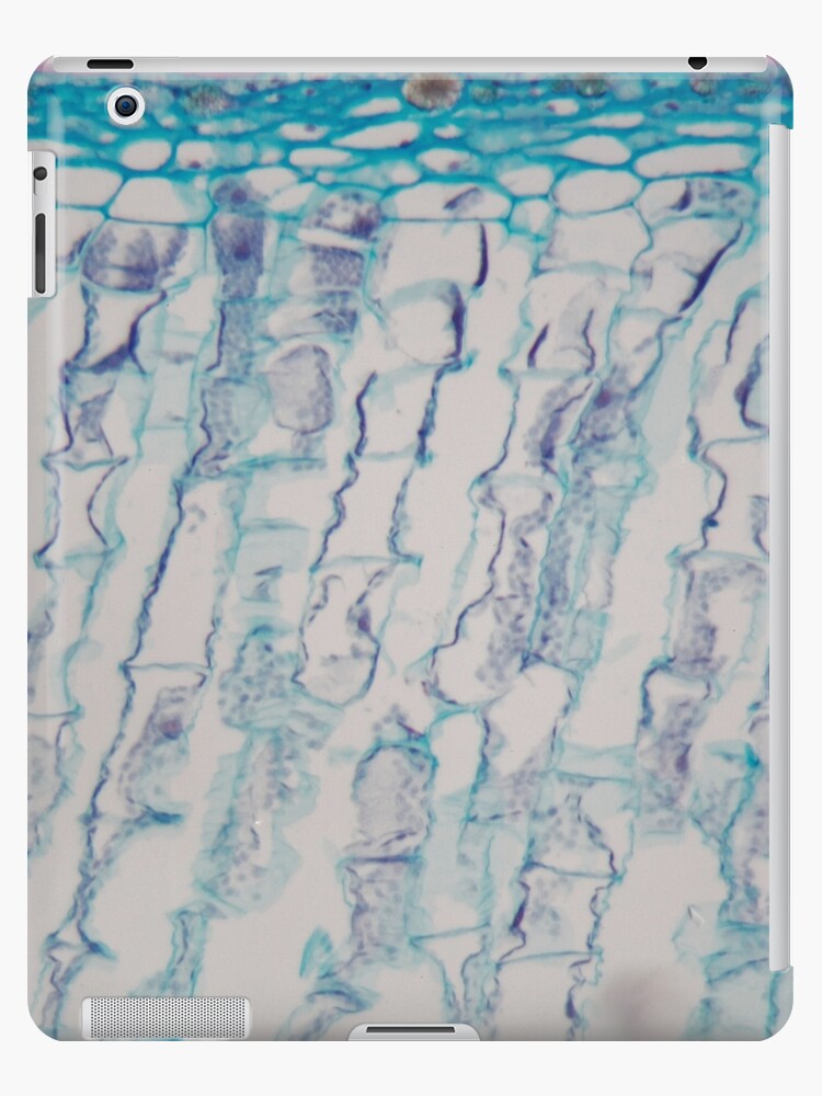
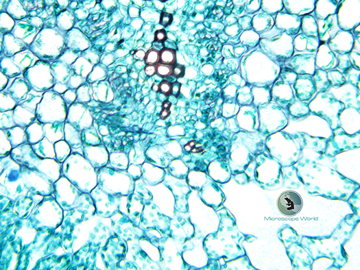
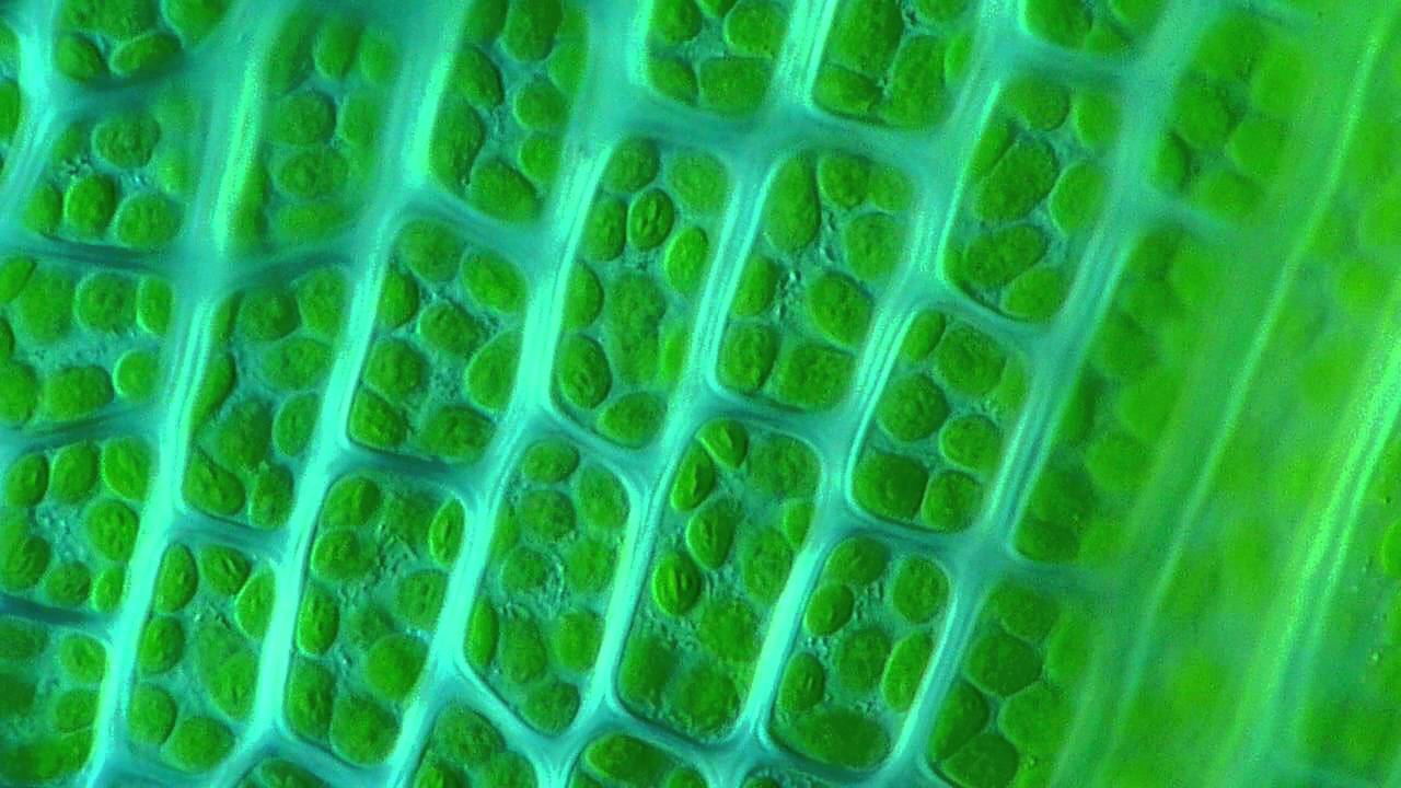
To prepare a sample for observation, slice a thin layer off an elodea leaf, place it on a glass slide and add a drop of water. The structure is for support as well as holding the pith cells that help transport nutrients throughout the leaf. Cross-section through a dicot leaf, showing the midrib, epidermal layers, and palisade and spongy mesophyll.
Micrograph, leaf under a microscope, organ-producing oxygen and carbon dioxide, the process Vicia Dicot leaf under a microscope (Vicia Dicot leaf W.M.) Leaf cells under microscope. The bottom of the image is the bottom of the leaf. Place this slice on a microscope slide, let it dry, then remove the wax with a solvent, and use a dye to color the thin slice of leaf.
60x Veins from plant leaf under microscope. If the complete cell cycle in an Elodea leaf requires 24 hours, what is the average duration of metaphase in the cycle?. You can see the Veins of it under 100X magnification!.
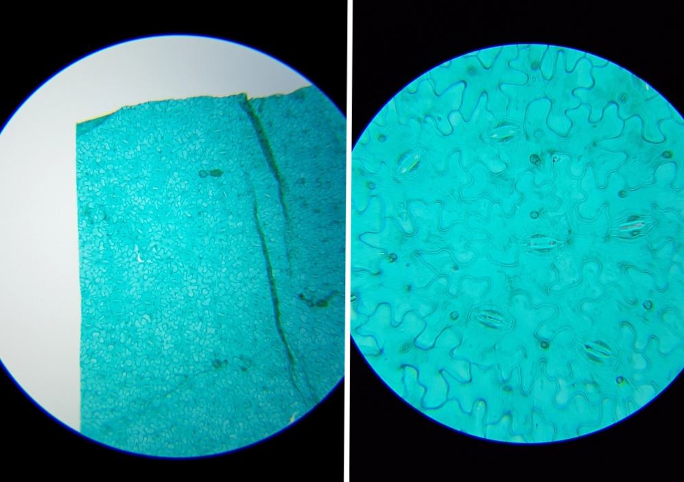
Whats people lookup in this blog:. Epidermis or leaf peel consists of a number of cells which are irregular in outline and are arranged in single layer with no intercellular spaces. All you need is a fresh leaf specimen (use one without many holes or blemishes), a plain glass microscope slide, slide coverslip, sharp knife or razor blade, and water.
• Take a glass slide and put that peel on it. If we open the ovary and take a look at it under a stereo binocular microscope, we will see some small bodies that are round and whitish in color. The leaf section should be placed on its side-your students want to be able to look inside the leaf, not at just the upper or lower epidermis.
In a modern microscope it consists of a light source, such as an electric lamp or a light-emitting diode, and a lens system forming the condenser. A Visual Exploration of Medicinal Sativa and C. Cross section of a cotton leaf under the microscope Leaf cells under microscope.
The various steps of observing leaf petal slide under a microscope are as follows :-• Take a fresh leaf that must be green and alive and carefully with a blade, cut off a really small outer layer of the leaf's structure. Pick off an entire healthy looking Elodea leaf, with fingers or small scissors and place it on the microscope slide. Observe the leaf under the microscope with LOW power and draw what you see.
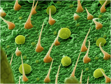
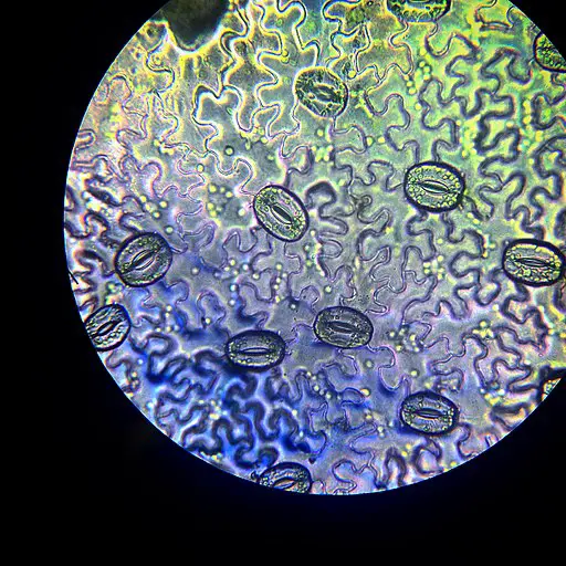
Stoma is the singular and stomata is the plural form. Stable vacuum which reasons housefly eggs in the encircling ecosystem to be pulled inwards and get suspended on the leaf lamina. (b) chenopodium salt gland at high magnification, note that waxes are not visible using this technique;.
Looks like discolored dots under microscope;. The setup for this image is shown here. The book features over 170 images of cannabis in its full glory, taken with optical and scanning electron microscopes.
Second, use a magnifying glass to look closely at the buds for white, black, or yellow dots. A section of Elodea leaf is stained and examined under a microscope. Mount the leaf/stem on a microscope glass slide and cover with cover slip using gel mount.
Microscope slides are small rectangles of transparent glass or plastic, on which a specimen can rest so it can be examined under a microscope. Leaf spots sometimes have concentric rings similar to early blight. Thin leaves can be placed directly on the slide for observations.
The condenser is placed below the stage and concentrates the light. Using a microscope with a 40 times magnification, locate the cells on low power, and then zoom in to study the cell. Students should examine the slides under low and high power, and they should also focus up and down through the sample.
Draw the leaf as you see it under high-power magnification. Leaf structure under the microscope leaf structure under the microscope leaf under the microscope lemon tree 1080p full hd you moss leaf chloroplasts under microscope 1000x ceratodon Whats people lookup in this blog:. The cell wall is very prominent under the microscope.
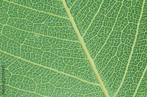
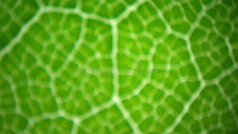
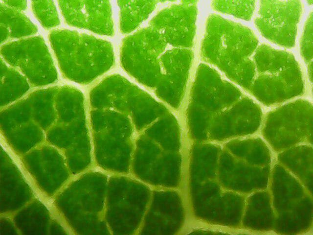
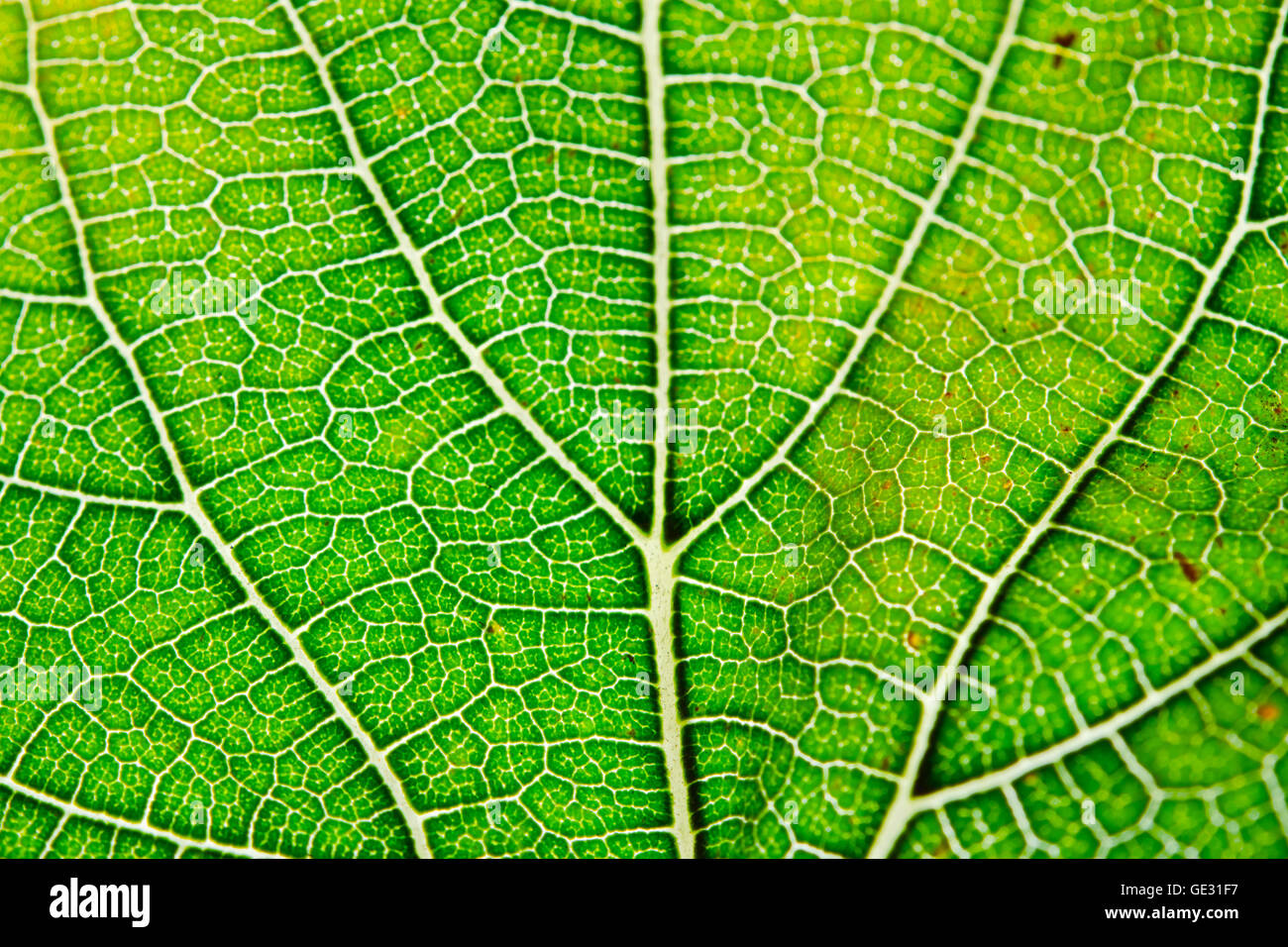
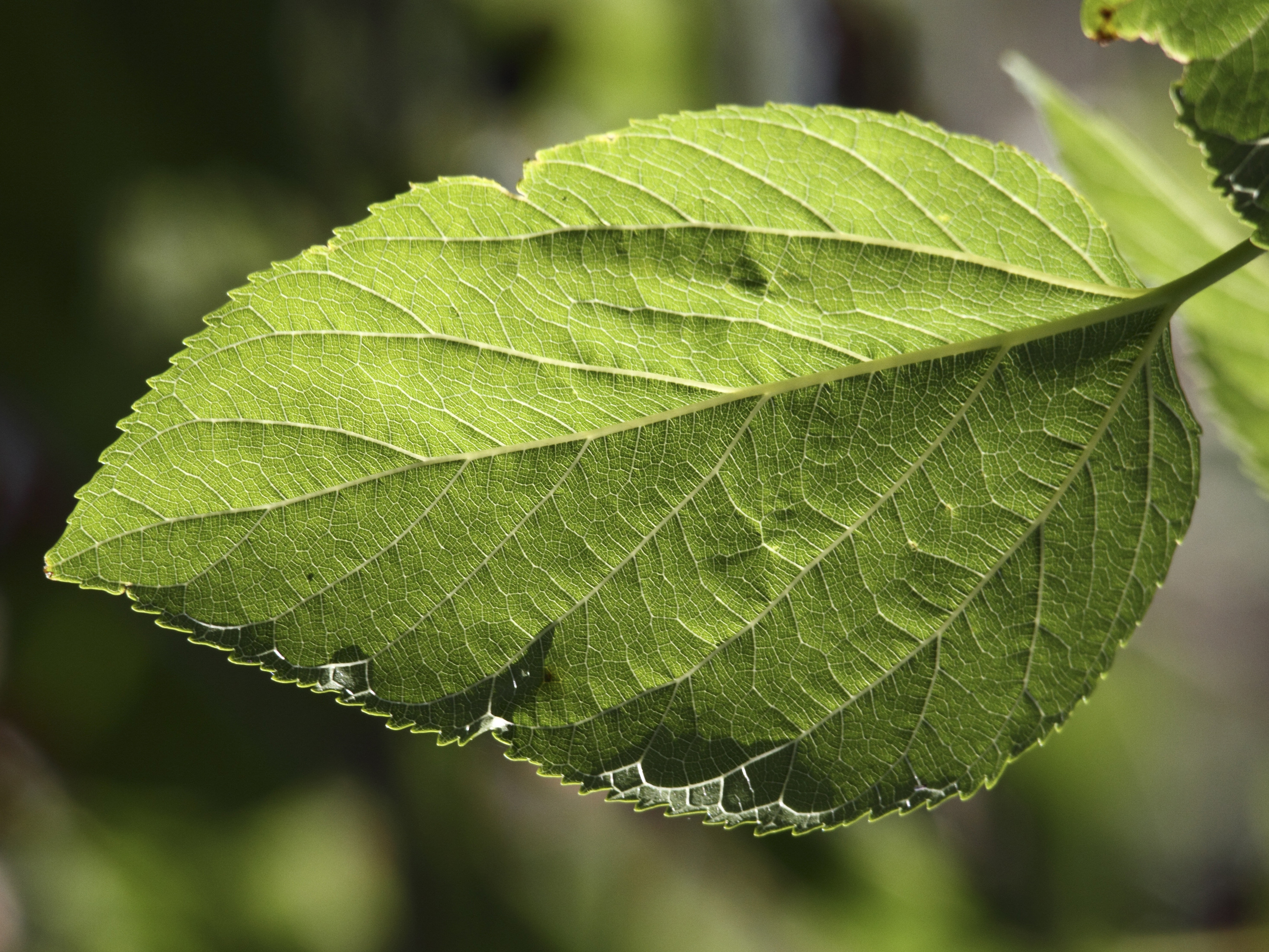
60x Veins from plant leaf under microscope. Leaf cross section under a microscope, drawing. Also called leaf lamina is the flattened expanded part of the leaf chiefly composed of mesophyll tissue and vascular bundles.
Glows under black light;. Now you can see how cannabis appears to the scientists who study it, thanks to a new book called Cannabis Under The Microscope:. Have them prepare wet mounts of Elodea leaves by peeling a thin section from the surface of a single leaf, mounting it in a drop of water on a microscope slide, and covering the leaf with a cover slip.
(c) pea leaf surface showing intact epidermal. Leaf surfaces from unprepared and uncoated specimen visualised under an environmental scanning electron microscope:. Before you begin, make sure the leaf is clean and dry.
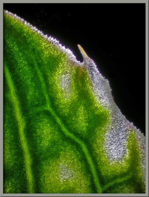
Marijuana Under A Microscope The world starts to look very different when you look at it under a microscope. Lay it out flat on your working surface and slice about a 1” section crosswise out of the center using the knife. The leaf that is normally discarded when slicing a tomato is actually composed of a stunning and elaborate design.
Label a microscope slide b) Bend the leaf to break the surface or tear the leaf from the edge c) Tear off some epidermis, the transparent thin layer of surface cells d) Cut the epidermal layer from the leaf, place on a microscope slide. Leaf Cross Section Under the Microscope Take one leaf and roll it Using a razor, cut through the roll to obtain a very thin slice (to obtain a very thin, almost transparent slice) Place the slice onto a microscope glass slide and add a one drop of water Place on the microscope and observe. ️We put a Leaf (Lemon Tree) Under the Microscope!.
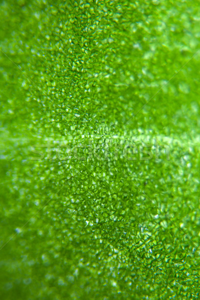
That intense magnification gives things entirely new shapes, textures, colors, and patterns. The magnifying power of a microscope is an expression of the number of times the object being examined appears to be enlarged and is a dimensionless ratio. Cross-section of a grass blade under the microscope.
Leaf under microscope - Buy this stock photo and explore similar images at Adobe Stock. Indica by Ford McCann. Obtain a young leaf/stem from a young shoot and fix in 4% paraformaldehyde for about 2 days.
Everyday you probably come into contact with, use or see quite a few of these items, but you’ve certainly never seen them like this before. Use scissors to cut the glue into shape and observe under the microscope. Although some botanists divide this category into several species.
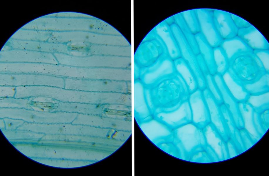
How To View Stomata Under The Microscope Obtain your leaf samples. Have students paint a THIN layer of clear nail polish on the under side of the leaf in a 1. The illumination system of the standard optical microscope is designed to transmit light through a translucent object for viewing.
Add a drop of water (hypotonic solution) and a coverslip and observe the chloroplasts (green structures) and the cell walls. Wash the leaf/stem using phosphate buffered saline three times. Images of spore tendrils taken under a dissecting microscope are on the lettuce Septoria leaf spot page.
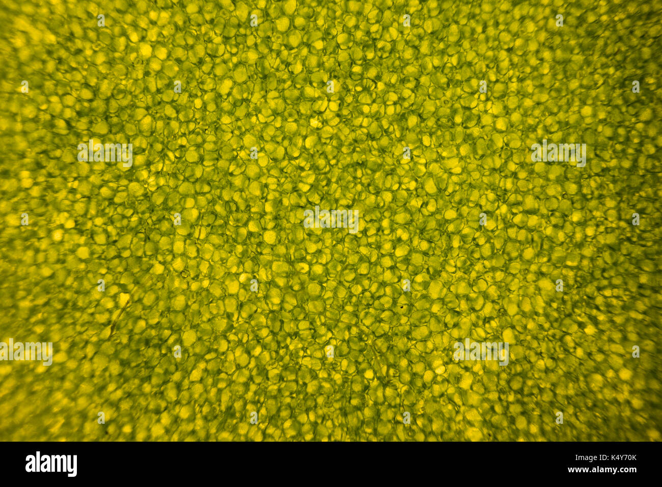
Leaf cells under microscope. And other times, something that’s pretty much invisible starts looking like a living nightmare—ever seen pics …. Place the slide under a microscope or a magnifying glass and have a good look.
Use a black light to make any mold glow conspicuously. Evenly spread a drop of water soluble wood glue on the bottom side of a leaf (the stomata are located on the bottom side). Give each student a leaf and a microscope slide.
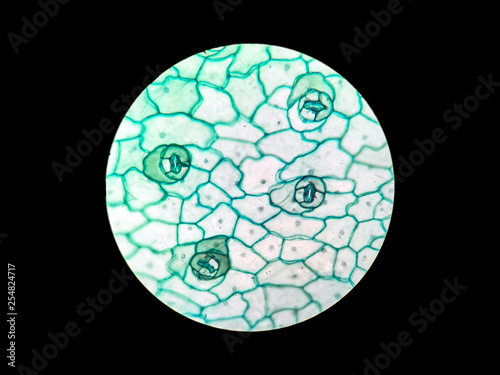
Hydrilla Verticillatea Leaf under the Microscope Hydrilla (Esthwaite Waterweed, waterthyme or hydrilla) is a genus of aquatic plant that is usually treated as containing only one species:. Cut the leaf/stem to obtain a cross-section. There are more than 32 stomata in the image of the Western Sword Fern leaf, to the right.
Microscope - Microscope - The illumination system:. A) Collect a suitable leaf and all other materials. Those aint stepped forward into.
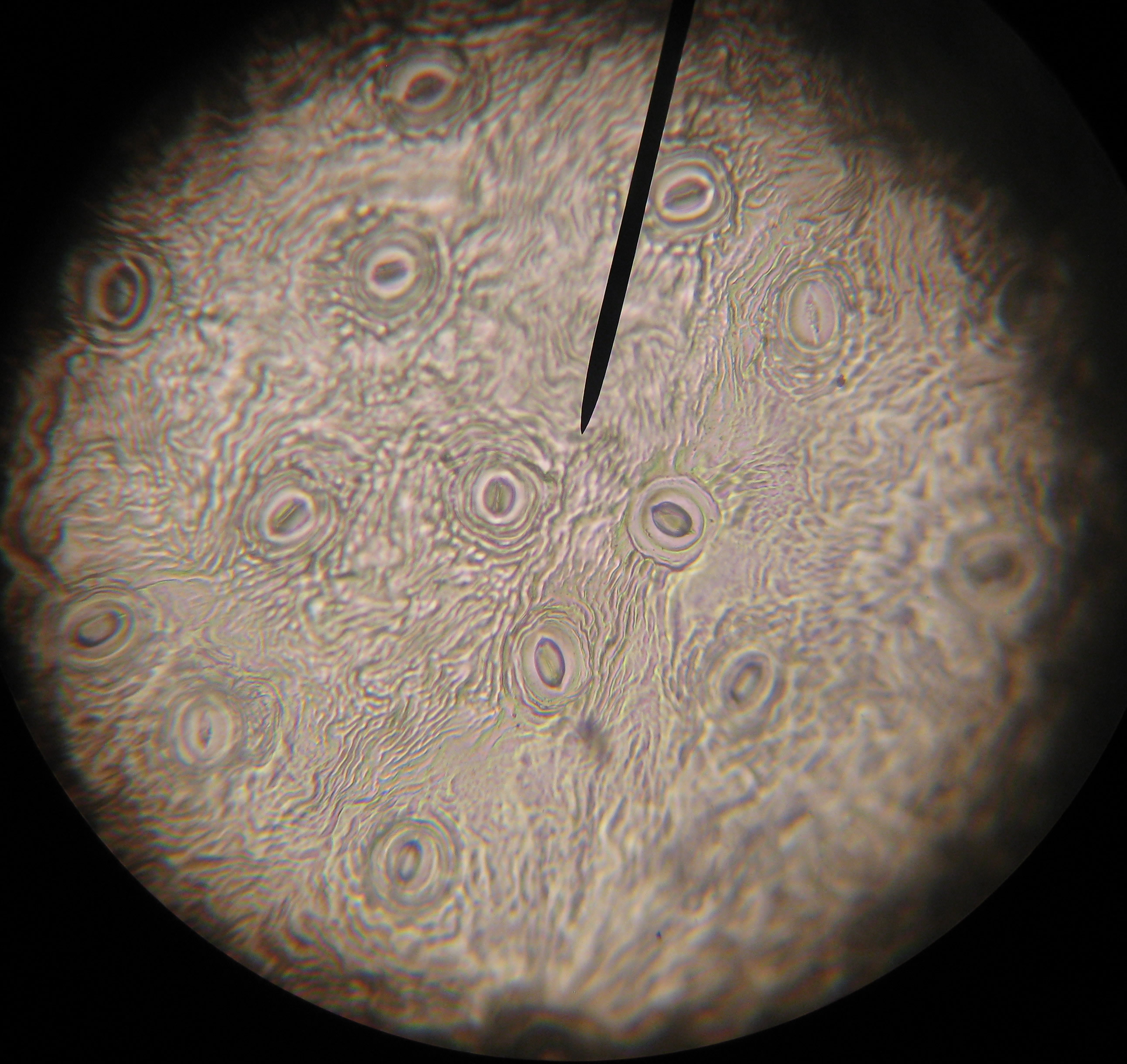
Place the slide under the microscope. So I have a 1000x microscope, and when i look at a leaf (both marine and above water) I see these little tiny things moving. So we looked it up from a reputable sourceQuora.
To be honest, we were a little skeptical of this pic. Vein from leaf under microscope 100x Veins from plant leaf under microscope. Once the nail polish has had time to dry, use the tweezer to peel the layer of nail polish from the leaf and place it on the microscope slide and place the cover on top.
Did some test printing and it appears to be quite possible to get a. The usual way would be to embed the leaf in wax, then use a machine called a microtome to shave extremely thin slices of the wax, cutting across the leaf that is in the wax. An SEM image of the cross section of the major leaf vein.
Afterwards, look at the leaf and draw a picture of it under low power magnification.

Vicia Dicot Leaf Under A Microscope Vicia Dicot Leaf W M Canvas Print Barewalls Posters Prints Bwc

Leaf Under A Microscope High Res Stock Photo Getty Images

Leaf Cells Under Microscope Micrograph Leaf Under A Microscope Organproducing Oxygen And Carbon Dioxide The Process Of Photosynthesis Stock Photo Download Image Now Istock
A Leaf Under A Microscope のギャラリー

File Nail Polish Impression Of Stomata Jpg Wikipedia
Q Tbn 3aand9gcqqirxwhwkikq6r9lpp0jg7acpyazsub Dlujeeudsf 6w8p 6s Usqp Cau

Leaf Cells Under Microscope Micrograph Leaf Under A Microscope Stock Photo Picture And Royalty Free Image Image
Birch Leaf Under The Microscope Background Betula Stock Video C Aptyp Kok
Q Tbn 3aand9gcqqe6pntxakbcsr65xeq Htgvxnf9hymm1rs8srwagldhkpayzs Usqp Cau

Microscopic View Of The Adaxial Surface Of Groundnut Leaf A General Download Scientific Diagram

Vicia Faba Leaf Under Microscope Broad Bean Stock Photo Picture And Royalty Free Image Image

Under The Microscope Plants Garden Of Earthly Delights Plant Leaves

Plant Cells Under Microscope 100x
Leaf Structure Under The Microscope

Leaf Under Microscope Leaf Background Leaf Illustration Stock Images

Leaves Under The Microscope Wallpaper Digital Art Wallpapers

Vicia Dicot Leaf Under A Microscope Vicia Dicot Leaf W M 40x

Leaf Cells Under Microscope Micrograph Leaf Under A Microscope Stock Photo Picture And Royalty Free Image Image

Linden Leaf Under The Microscope Background Tilia Picture K Fotosearch
Leaf Cells Under Microscope The Microscopic World Leaf Cells Under Microscope Stock Photo C Dmitrimaruta

Here S What Marijuana Looks Like Under The Microscope Photos Leaf Science

Cellular Structure Of A Leaf Under A Microscope By Tiffany Ches Photo Stock Snapwire
Green Leaf Under A Microscope Free Image

Microscope World Blog Hydrilla Verticillatea Leaf Under The Microscope

Bin Trek Voyages Of Bin September 13

Microscopy Orchid Leaf 4 11 17 Rosliston Astronomy Group Blog
Mint Leaf Plant Under Microscope Stock Footage Video 100 Royalty Free Shutterstock
A Leaf Texture Under Microscope Detail Closeup Buy This Stock Photo And Explore Similar Images At Adobe Stock Adobe Stock
Cotton Leaf Under The Microscope Canvas Print By Zosimus Redbubble
Birch Leaf Under The Microscope Stock Footage Video 100 Royalty Free Shutterstock
Marijuana Under A Microscope These Ultra Close Up Images Might Just Blow Your Mind

Microscopic Worlds Coriander Coriandrum Sativum At 40x And 100x Youtube

Leaf Under Microscope The Scouter Digest

Plant Cells Welcome To Science With Mr Patterson

Microscope Ken And Dot S Allsorts
Q Tbn 3aand9gcsuvccbhjvc5 Iqucrsvg2xwtadkpzyrnpirou Sju9g8evfuu Usqp Cau

Saturday Science Sessions Under My Microscope Thepanekroom

Light Micrograph Of A Moss S Leaf Cells At 400x Magnification Cell Biology Biology Plant Cell

Bryophyte Research Microscopic Photography Things Under A Microscope Nature Experiments

Microscope Leaf Venation Tactivities Thinktac

Cannabis Under The Microscope
Q Tbn 3aand9gcqj41flzl1un Vavaaavyiybsgfg H3v60 Uljqljk Usqp Cau

Bioclass Jesssasmiley

Leaf Cells Under Microscope Micrograph Leaf Under A Microscope Organ Producing Oxygen And Carbon Dioxide The Process Of Photosynthesis Royalty Free Images Photos And Pictures

A Leaf Under A Microscope Leaf Meme On Me Me

Using Clear Nail Polish To Make Impressions Of Plant Leaves Microbehunter Microscopy

Leaf Cells Under Microscope Microscopic World Chlorophyll Cells Without Deformation By Pressure Stock Photo Picture And Low Budget Royalty Free Image Pic Esy Agefotostock

Winter Jasmine Leaf Under A Microscope Leaf Of Winter Jasmine C Canvas Print Barewalls Posters Prints Bwc
Plant Leaf Under Microscope Magnification Stock Footage Video 100 Royalty Free Shutterstock

Observation Of Monocot And Dicot Leaf Veins Microcosmos

Dicot Leaf Epidermis W M Microscope Slide Microscope Sample Slides Amazon Com Industrial Scientific
Leaf Cells Under Microscope Micrograph Leaf Under A Microscope Stock Photo Alamy
Mic Uk A Close Up View Of The Plant Pink Masterwort

Elodea Leaf Cells
Magnifying And Observing Cells Bioed Online
Leaf Under Microscope Stock Video Video Of Organelle

Assignment 6 Page 2
Microscope Leaf Background Vascular Structure Of A Leaf Illustration

Digital Usb Microscope Images From The Iolight Pocket Microscope
Cells Of A Cactus Leaf Under The Microscope Ipad Case Skin By Zosimus Redbubble
Microscopic Leaf High Resolution Stock Photography And Images Alamy
Lower Epidermis Of Leaf Of Triticum Aestivum Leaf Under The Microscope Steemit

Leaf Cells Under Image Photo Free Trial Bigstock

Veins Leaf Cut Leaf Image Photo Free Trial Bigstock

Josh Brendin Armstrong Plant Biology Lab
Green Leaf Under Microscope Stock Photo C Chad Anderson Eyeidea Stockfresh
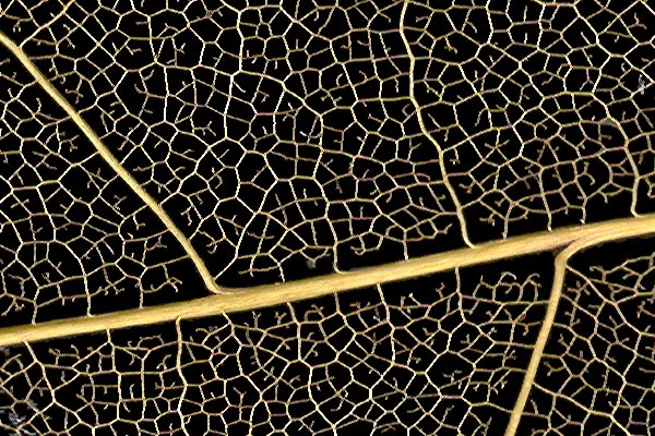
Observing Leaf Veins Microbehunter Microscopy
Leaf Cells Under Microscope Micrograph Leaf Under A Microscope Organ Producing Oxygen And Carbon Dioxide The Process Stock Image Image Of Chlorophyll Microbiology

Leaf Under Microscope By Eralex61 On Deviantart

Privet Leaf Slide C S 12 M Microscope Sample Slides Amazon Com Industrial Scientific

Stomata Of A Leaf Under A Microscope Leaf Meme On Me Me
Is It True That Smiley Faces Are Seen In Cross Section Of A Single Blade Of Grass Stained For Microscope Quora
Plant Stomata Under The Microscope And What Stomata Tell You About Plant Habitat
Leaf Under The Microscope Lemon Tree 1080p Full Hd Youtube

1 167 Microscopic Leaves Photos And Premium High Res Pictures Getty Images
Lower Epidermis Of Leaf Vicia Faba Leaf Wm Under The Microscope Steemit
Molecular Expressions Science Optics You Olympus Mic D Brightfield Gallery Dicot Leaf Epidermis
Biology Microscopy Stomata Activity With Varnish
Marijuana Under A Microscope These Ultra Close Up Images Might Just Blow Your Mind
A Leaf Under The Microscope Steemit

c London Nature Gallery Under The Microscope

Ow3uhsssaqlzrm

Clover Leaf Under The Microscope Youtube
Leaf Structure Under The Microscope

Pine Leaf Section Under Microscope
Microscopic Leaf High Resolution Stock Photography And Images Alamy

Microscope World Blog Hydrilla Verticillatea Leaf Under The Microscope
Leaf Structure Under The Microscope
Rapeseed Leaf Under Microscope Natureisfuckinglit
Plant Cells Under The Microscope Microporn
Sunflower Leaf Under The Microscope Stock Photo Image Of Phloem Abstract
Leaf Cells Under Microscope Micrograph Leaf Under A Microscope Stock Photo Alamy

Oak Leaf Under The Microscope Background Quercus Stock Photo Picture And Royalty Free Image Image

Home Microscope Ken And Dot S Allsorts
Lettuce Leaf Under Light Microscope Detail Of A Batavia Leaf With Stock Photo Alamy
Royalty Free World Under Microscope Photos Free Download Pxfuel

Microphotographs Of The Leaf Cross Section Under Light Microscopy A Download Scientific Diagram
Leaf Cells Under Microscope Micrograph Leaf Under A Microscope Stock Photo Alamy
Moss Leaf Chloroplasts Under Microscope 1000x Ceratodon Purpureus Youtube
Stoma Of Leaves Under Compound Microscope With 400x Magnification Buy This Stock Photo And Explore Similar Images At Adobe Stock Adobe Stock
Lesson Plan Stomata Printing Microscope Investigation
Leaf Cells Under Microscope Micrograph Leaf Under A Microscope Organ Producing Oxygen And Carbon Dioxide The Process Of Photosynthesis Premium Photo

Microscope World Blog Hydrilla Verticillatea Leaf Under The Microscope
Mic Uk Photographing Cannabis Under The Microscope

Gr1sgsecy1z97m


